

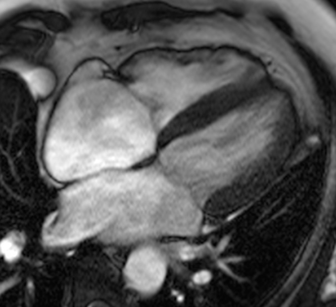
Understanding why cardiac MRI is done helps patients make informed decisions about their heart health. Cardiac magnetic resonance imaging (MRI) is a sophisticated, non-invasive imaging technique that provides detailed pictures of the heart’s structure and function, helping Cardiologists diagnose various heart conditions with exceptional precision.
Dr Navin Chandra, an experienced Cardiologist practising in Berkshire (Windsor, Ascot, Slough & surrounding areas) specialises in cardiovascular imaging and Interventional Cardiology. With years of experience in cardiac imaging, PCI, and TAVI procedures, Dr Chandra uses advanced diagnostic techniques to provide comprehensive heart care for patients across the region.
What Is Cardiac MRI and How Does It Work?
Cardiac MRI uses powerful magnetic fields and radio waves to create detailed images of your heart without using radiation. Unlike other imaging methods, cardiac MRI can capture both still images and moving pictures of your heart as it beats, providing valuable information about heart function and blood flow.
The procedure typically takes 30-60 minutes and involves lying on a table that slides into a large, tube-shaped machine. You may receive a contrast agent through an intravenous line to enhance image quality and help identify specific heart conditions more clearly.
Why Is Cardiac MRI Done for Heart Conditions?
Cardiac MRI is recommended for various reasons, each providing crucial information about your heart health. The detailed images help Cardiologists evaluate heart structure, function, and blood flow with remarkable accuracy.
Assessing Heart Function and Structure
Cardiologists use cardiac MRI to evaluate how well your heart pumps blood and examine the heart’s chambers, walls, and valves. This comprehensive assessment helps identify:
Heart muscle weakness or damage
Abnormal heart chamber sizes
Problems with heart valve function
Congenital heart defects
Heart muscle abnormalities
Diagnosing Heart Muscle Disease
Cardiac MRI excels at detecting various heart muscle conditions that may not be visible on other imaging tests. The scan can identify inflammation, scarring, or infiltration of the heart muscle, helping diagnose conditions such as cardiomyopathy, myocarditis, and cardiac amyloidosis.
Evaluating Blood Flow and Coronary Arteries
The imaging technique can assess blood flow through the heart and major blood vessels, identifying areas where blood flow may be reduced or blocked. This information proves valuable for planning treatments such as PCI or other interventional procedures.
When Might Your Cardiologist Recommend Cardiac MRI?
Several symptoms and conditions may prompt your Cardiologist to recommend cardiac MRI as part of your diagnostic evaluation.
Unexplained Chest Pain or Breathlessness
If you experience chest pain or shortness of breath that cannot be explained by initial tests, cardiac MRI may help identify the underlying cause. The detailed images can reveal heart problems that might not be detected through other diagnostic methods.
Abnormal Heart Rhythm Investigations
For patients with irregular heartbeats or arrhythmias, cardiac MRI can identify structural heart problems that may be contributing to rhythm disturbances. This information helps guide treatment decisions and assess the need for specific interventions.
Follow-up After Heart Attack
Following a heart attack, cardiac MRI provides detailed information about the extent of heart muscle damage and helps assess recovery. The scan can identify viable heart muscle and areas of scarring, informing treatment planning and prognosis.
What Are the Benefits of Cardiac MRI?
Cardiac MRI offers several advantages over other heart imaging techniques, making it a valuable diagnostic tool for comprehensive heart evaluation.
No radiation exposure, making it safer for repeated examinations
Excellent image quality with detailed soft tissue contrast
Ability to assess both heart structure and function in one examination
Non-invasive procedure with minimal patient discomfort
Provides information about blood flow and heart muscle viability
Can detect early signs of heart disease before symptoms develop
Preparing for Your Cardiac MRI
Proper preparation ensures the best possible images and a smooth examination experience. Your Cardiologist will provide specific instructions based on your individual circumstances.
Before your scan, inform your healthcare team about any metal implants, pacemakers, or claustrophobia concerns. You may need to avoid eating for several hours before the examination, particularly if contrast material will be used. Comfortable, metal-free clothing is recommended for the procedure.
Understanding Your Cardiac MRI Results
After your cardiac MRI, a specialist Cardiologist will analyse the images and provide a detailed report. Dr Chandra will then discuss the results with you, explaining what the findings mean for your heart health and any recommended next steps.
The results may show normal heart function, identify specific heart conditions requiring treatment, or provide information to guide further investigations. Dr Chandra will use these findings alongside your symptoms and other test results to develop an appropriate treatment plan.
Conclusion
Cardiac MRI is done to provide detailed, accurate information about your heart’s structure and function, helping Cardiologists diagnose and monitor various heart conditions. This advanced imaging technique offers valuable insights that guide treatment decisions and improve patient outcomes.
If you have concerns about your heart health or have been recommended for cardiac imaging, Dr Navin Chandra provides expert cardiac MRI services in Berkshire (Windsor, Ascot, Slough & surrounding areas). To discuss whether cardiac MRI may be suitable for your situation, book a consultation with Dr Navin Chandra today.
Have Questions About Your Heart Health?
Schedule a consultation with Dr Chandra to discuss personalized strategies for maintaining optimal cardiovascular health.


