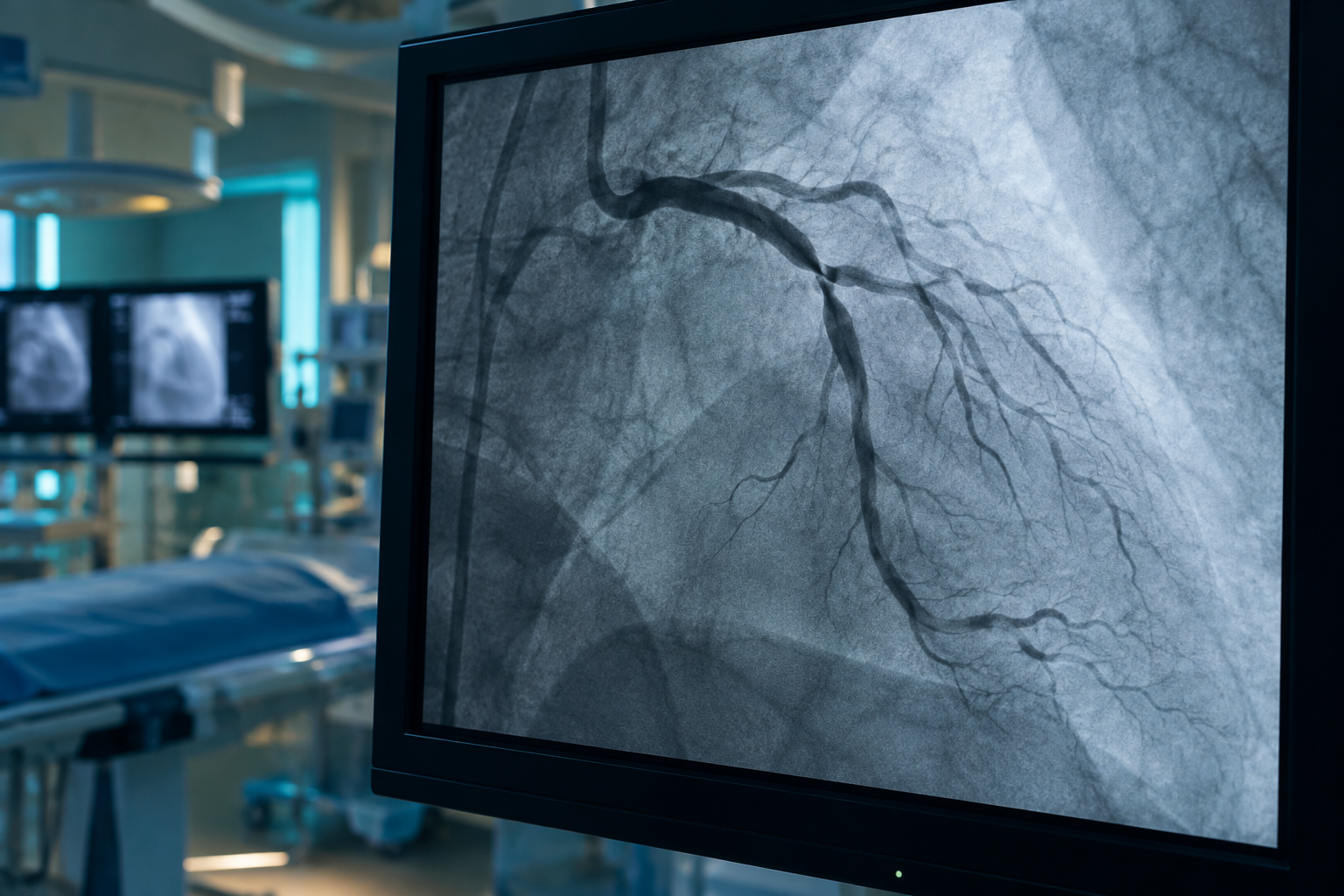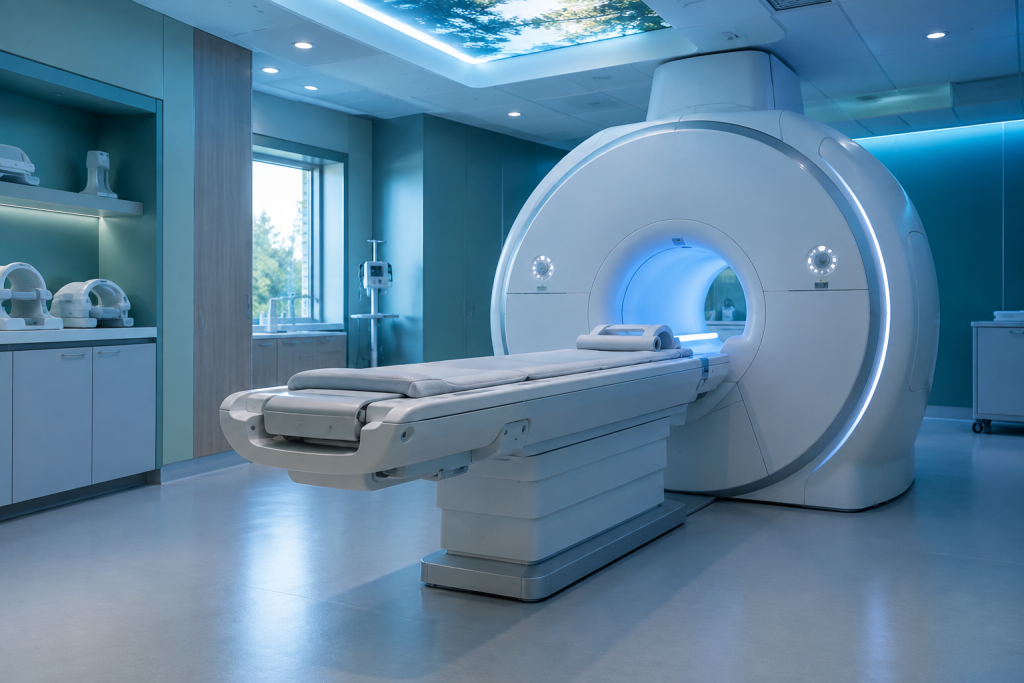
Understanding how cardiac MRI works is essential for patients undergoing this advanced imaging technique. Cardiac MRI (Magnetic Resonance Imaging) is a non-invasive tool that provides detailed images of the heart and surrounding structures, helping in the diagnosis and management of various cardiac conditions. Dr Navin Chandra, a Consultant Cardiologist with extensive training and experience in cardiovascular health, employs this technology to enhance patient outcomes at his practice in East Berkshire & South Buckinghamshire.
What is Cardiac MRI?
Cardiac MRI is a state-of-the-art imaging modality that uses magnetic fields and radio waves to create detailed images of the heart. Unlike traditional X-rays or CT scans, cardiac MRI does not involve ionising radiation, making it a safer option for many patients.
This imaging technique is particularly useful for evaluating heart structures, assessing blood flow, and identifying conditions such as myocardial infarction, cardiomyopathy, and congenital heart defects.
How Does Cardiac MRI Work?
Cardiac MRI works by utilising powerful magnets and radiofrequency pulses to generate images of the heart. Here’s a simplified breakdown of the process:
- Patient Preparation: Before the procedure, patients may be asked to refrain from eating or drinking for a few hours. They will also be required to remove any metal objects, such as jewellery, that could interfere with the MRI.
- Positioning: Patients lie on a movable table that slides into the MRI machine. They must remain still during the imaging process to ensure clear images.
- Imaging Process: As the MRI machine generates images, patients may hear a series of loud tapping or thumping noises. The technician may instruct them to hold their breath for short periods to capture clear images of the heart.
- Contrast Agents: In some cases, a contrast agent may be injected into a vein to enhance the images. This helps to better visualise blood flow and any abnormalities in the heart tissue.
- Image Analysis: After the imaging is complete, a MRI specialist Radiologist or Cardiologist will analyse the images to identify any issues.
What Conditions Can Cardiac MRI Diagnose?
Cardiac MRI is instrumental in diagnosing a variety of cardiac conditions, including:
- Coronary Artery Disease: It helps in assessing the extent of damage to the heart muscle due to reduced blood flow.
- Cardiomyopathy (heart muscle abnormalities): Cardiac MRI can evaluate the size and function of the heart chambers, providing insights into the type of cardiomyopathy.
- Heart Valve Disease: This imaging can help in the assessment of heart valves, detecting any abnormalities.
- Congenital Heart Defects: It is useful in visualising structural heart defects present from birth.
- Myocarditis: Cardiac MRI can indicate inflammation of the heart muscle, aiding in diagnosis.
What Are the Benefits of Cardiac MRI?
Cardiac MRI offers several advantages over other imaging modalities:
- No Radiation Exposure: As mentioned earlier, cardiac MRI does not use ionising radiation, making it a safer option for patients, especially those requiring multiple scans.
- High-Resolution Images: MRI provides detailed images that can reveal subtle abnormalities that other imaging techniques might miss. Studies have shown that cardiac MRI can detect myocardial infarction with an accuracy of up to 95%.
- Functional Information: It can assess not just the structure of the heart, but also its function, including blood flow and pumping efficiency.
- Versatility: Cardiac MRI can be adapted to evaluate various conditions, from coronary artery disease to congenital heart defects.
What to Expect During a Cardiac MRI?
Patients may feel anxious about undergoing a cardiac MRI. Knowing what to expect can help alleviate some of these concerns:
- Duration: The procedure typically takes between 30 to 90 minutes, depending on the complexity of the images required.
- Comfort: The MRI machine is enclosed, which can be challenging for those with claustrophobia. Patients can discuss this with their clinician beforehand to explore options for comfort.
- Post-Procedure: After the MRI, patients can usually resume their normal activities immediately, unless advised otherwise.
Are There Any Risks Associated with Cardiac MRI?
Cardiac MRI is generally safe, but there are some considerations:
- Metal Implants: Patients with certain metal implants, such as pacemakers or certain types of stents, may not be eligible for an MRI. It is crucial to inform the medical team about any implants before the procedure.
- Contrast Reactions: While rare, some patients may experience allergic reactions to the contrast agent used during the MRI.
- Kidney Function: Patients with compromised kidney function should discuss the use of contrast agents with their clinician, as it may pose risks.
How to Prepare for a Cardiac MRI?
Preparation for a cardiac MRI is quite straightforward:
- Inform Your Doctor: Discuss any medical history, allergies, and current medications with your clinician before the MRI.
- Dress Comfortably: Wear loose-fitting clothing without metal fasteners. You may be provided with a gown to wear during the procedure.
- Follow Fasting Instructions: If required, adhere to any fasting instructions given by your healthcare provider.
What Happens After a Cardiac MRI?
Once the MRI is completed, your healthcare provider will review the images. A follow-up appointment may be scheduled to discuss the results and any necessary treatment options. Depending on the findings, additional tests or interventions may be recommended to address any identified issues.
Frequently Asked Questions
How does cardiac MRI work?
Cardiac MRI works by using strong magnetic fields and radio waves to generate detailed images of the heart, helping to diagnose various heart conditions without the use of ionising radiation.
Is cardiac MRI safe?
Yes, cardiac MRI is generally safe and does not involve radiation. However, patients with certain metal implants should consult their doctor before undergoing the procedure.
What should I expect during the procedure?
During a cardiac MRI, you will lie on a table that slides into the MRI machine. You may hear loud noises and will need to remain still for short periods. The procedure typically lasts between 30 to 90 minutes.
Are there any side effects from the contrast agent used?
While most patients tolerate the contrast agent well, some may experience allergic reactions. It’s essential to inform your clinician of any known allergies prior to the procedure.
How long does it take to get results from a cardiac MRI?
Results from a cardiac MRI can usually be available within a few days, but this timeframe may vary based on the complexity of the images and the workload of the imaging centre.
Can I eat before a cardiac MRI?
In most cases, you may be advised to avoid eating or drinking for a few hours before the procedure, especially if a contrast agent is to be used. Always follow your healthcare provider’s instructions.
To find out whether how cardiac MRI works may be suitable for you, book a consultation with Dr Navin Chandra, a specialist in cardiovascular health. His expertise ensures comprehensive management of all adult cardiac conditions, from prevention through advanced intervention.
Have Questions About Your Heart Health?
Schedule a consultation with Dr Chandra to discuss personalized strategies for maintaining optimal cardiovascular health.