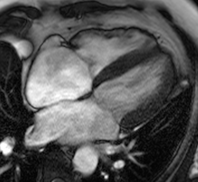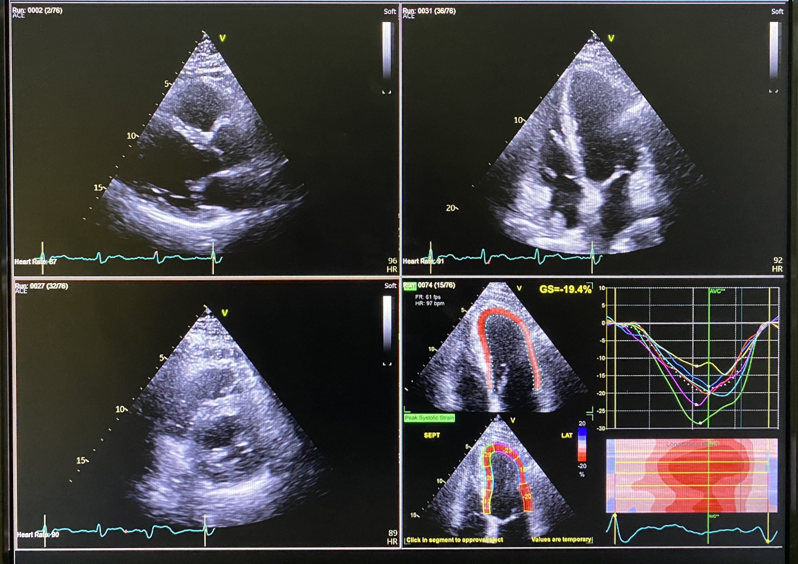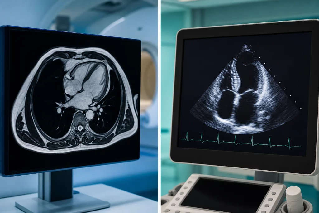
Whether cardiac MRI is better than echo depends on your specific heart condition and what your Cardiologist needs to investigate. Both cardiac magnetic resonance imaging (MRI) and echocardiography (echo) are valuable diagnostic tools that provide different types of information about your heart’s structure and function.
Dr Navin Chandra, a Consultant Cardiologist practising in Berkshire (Windsor, Ascot, Slough & surrounding areas), specialises in cardiovascular imaging and uses both techniques to diagnose and monitor various heart conditions. His expertise in cardiac imaging helps patients receive the most appropriate scan for their individual circumstances.
What Is the Difference Between Cardiac MRI and Echo?
Echocardiography uses ultrasound waves to create real-time images of your heart, whilst cardiac MRI uses powerful magnetic fields and radio waves to produce detailed cross-sectional images. Each technique offers distinct advantages depending on what your Cardiologist needs to examine.
An echo scan typically takes 30-45 minutes and can be performed in most clinical settings. The procedure involves placing a small probe on your chest that sends sound waves through your body to create moving images of your heart beating.
Cardiac MRI scans usually take 45-90 minutes and require specialised equipment. You’ll lie inside a large tube-shaped scanner whilst detailed images are captured from multiple angles, providing comprehensive views of your heart’s structure and blood flow.
When Is Cardiac MRI Better Than Echo?
Cardiac MRI may be recommended when your Cardiologist needs extremely detailed images of your heart’s structure or when echo results are inconclusive. This advanced imaging technique excels in several specific situations.
Complex Heart Conditions
For complex congenital heart disease, cardiac MRI provides superior detail of heart structures and blood vessels. The scan can accurately measure chamber sizes, wall thickness, and identify abnormal connections between heart chambers that may not be clearly visible on echo.
Tissue Characterisation
Cardiac MRI can distinguish between different types of heart tissue, including scar tissue from previous heart attacks. This capability makes it invaluable for assessing conditions like cardiomyopathy, where the heart muscle becomes diseased or damaged.
Precise Measurements
When exact measurements of heart function are crucial, cardiac MRI offers superior accuracy. This precision is particularly important for monitoring conditions over time or making treatment decisions for procedures like valve replacement.
When Is Echo Better Than Cardiac MRI?
Echocardiography remains the first-line imaging choice for many heart conditions due to its accessibility, speed, and excellent diagnostic capabilities for common cardiac problems.
Valve Assessment
Echo excels at evaluating heart valve function, showing how well valves open and close in real-time. The technique can accurately measure blood flow through valves and detect conditions like valve stenosis or regurgitation.
Emergency Situations
In urgent cardiac situations, echo provides immediate results without the time constraints of MRI scheduling. The portable nature of echo equipment means it can be performed at your bedside if necessary.
Routine Monitoring
For ongoing monitoring of known heart conditions, echo offers a convenient and cost-effective solution. Regular echo scans can track changes in heart function without the complexity of repeated MRI procedures.
What Are the Limitations of Each Scan?
Understanding the limitations of both cardiac MRI and echo helps explain why your Cardiologist might recommend one over the other for your specific situation.
Echo limitations include reduced image quality in patients with certain body types, lung disease, or chest wall abnormalities. Some heart structures may be difficult to visualise clearly, particularly the back wall of the heart or areas obscured by ribs.
Cardiac MRI limitations include longer scan times, higher costs, and contraindications for patients with certain metal implants or severe claustrophobia. The technique also requires patients to hold their breath multiple times during the scan, which some people find challenging.
How Does Your Cardiologist Choose Between Cardiac MRI and Echo?
Your Cardiologist considers several factors when determining whether cardiac MRI is better than echo for your particular case. These include your symptoms, medical history, previous test results, and the specific information needed to guide your treatment.
Initial cardiac assessment often begins with echo due to its accessibility and comprehensive overview of heart function. If echo results are normal and explain your symptoms, further imaging may not be necessary.
However, if echo findings are unclear, incomplete, or suggest complex pathology, your Cardiologist may recommend cardiac MRI for additional detail. Sometimes both scans provide complementary information that helps create a complete picture of your heart health.
What Should You Expect During Each Procedure?
Both cardiac MRI and echo are non-invasive procedures, but the experience differs significantly between the two scans.
During an echo, you’ll lie on your side whilst gel is applied to your chest. The sonographer moves a probe across your chest to capture images from different angles. You may be asked to change positions or breathe in specific ways to improve image quality.
For cardiac MRI, you’ll lie on a moveable table that slides into the scanner. You’ll need to remain still and may be asked to hold your breath for short periods. The scanner makes loud knocking sounds, so you’ll be given ear protection or headphones.
Are There Any Risks or Side Effects?
Both cardiac MRI and echo are considered very safe procedures with minimal risks for most patients.
Echo has no known risks or side effects as it uses sound waves rather than radiation. The procedure is safe for pregnant women and can be repeated as often as necessary without health concerns.
Cardiac MRI is also very safe for most people, though it’s not suitable for patients with certain metal implants, pacemakers, or severe kidney disease. Some patients may feel claustrophobic during the scan, but sedation can be provided if needed.
If you’re considering cardiac imaging to investigate heart symptoms or monitor a known condition, Dr Navin Chandra can help determine whether cardiac MRI or echo would be most appropriate for your situation. To discuss your cardiac imaging options and book a consultation, please contact Dr Navin Chandra’s practice in Berkshire.
Have Questions About Your Heart Health?
Schedule a consultation with Dr Chandra to discuss personalized strategies for maintaining optimal cardiovascular health.