Most people require a range of different tests to investigate their risk profile, understand the cause of symptoms and evaluate for a potential diagnosis of heart disease. Broadly speaking, these tests that can be categorised as follows:
- Bedside tests (e.g. blood tests or 12-lead ECG)
- Physiological tests (e.g. heart rhythm monitoring, blood pressure monitoring or exercise stress testing)
- Heart imaging tests (e.g. echocardiography, CT coronary angiography or cardiac MRI imaging)
- Invasive diagnostic tests (e.g. Reveal device implantation or coronary angiography)
Each of these tests investigate different facets of your heart health and provide invaluable diagnostic information. Dr Chandra will explain each test you require in detail so you understand why it is required and what information we hope to gain.
Blood Testing:
It Is often used to assess your cardiovascular risk factor profile, to evaluate for causes of your symptoms or to monitor your response to treatment. A blood sample is taken with a small needle from a vein in the arm. The equipment used is sterile and ‘single-use’ only. The sample is sent for analysis in a laboratory and results are available within a few hours. Routinely we will test your blood count, electrolyte levels, kidney & liver function, cholesterol & glucose levels. We may also test for specific heart muscle proteins (BNP, troponin), blood clotting levels and some hormone levels.
It Is often used to assess your cardiovascular risk factor profile, to evaluate for causes of your symptoms or to monitor your response to treatment. A blood sample is taken with a small needle from a vein in the arm. The equipment used is sterile and ‘single-use’ only. The sample is sent for analysis in a laboratory and results are available within a few hours. Routinely we will test your blood count, electrolyte levels, kidney & liver function, cholesterol & glucose levels. We may also test for specific heart muscle proteins (BNP, troponin), blood clotting levels and some hormone levels.

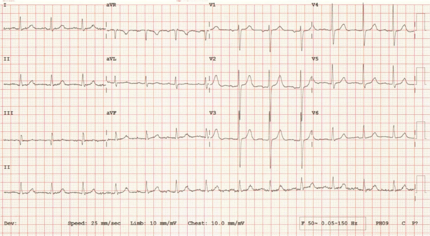
12-Lead ECG:
It Is a non-invasive and painless test to evaluate the electrical activity of your heart. This takes a few minutes to perform but requires you to remove clothing from the chest. ECG stickers are placed on the chest, arms and legs and connected to an ECG machine. You will need to lie still for an accurate recording to be made and printed onto a page. This simple test provides a snapshot of the electrical activity, structure and function and even blood supply of the heart and is useful to guide other investigations.
It Is a non-invasive and painless test to evaluate the electrical activity of your heart. This takes a few minutes to perform but requires you to remove clothing from the chest. ECG stickers are placed on the chest, arms and legs and connected to an ECG machine. You will need to lie still for an accurate recording to be made and printed onto a page. This simple test provides a snapshot of the electrical activity, structure and function and even blood supply of the heart and is useful to guide other investigations.
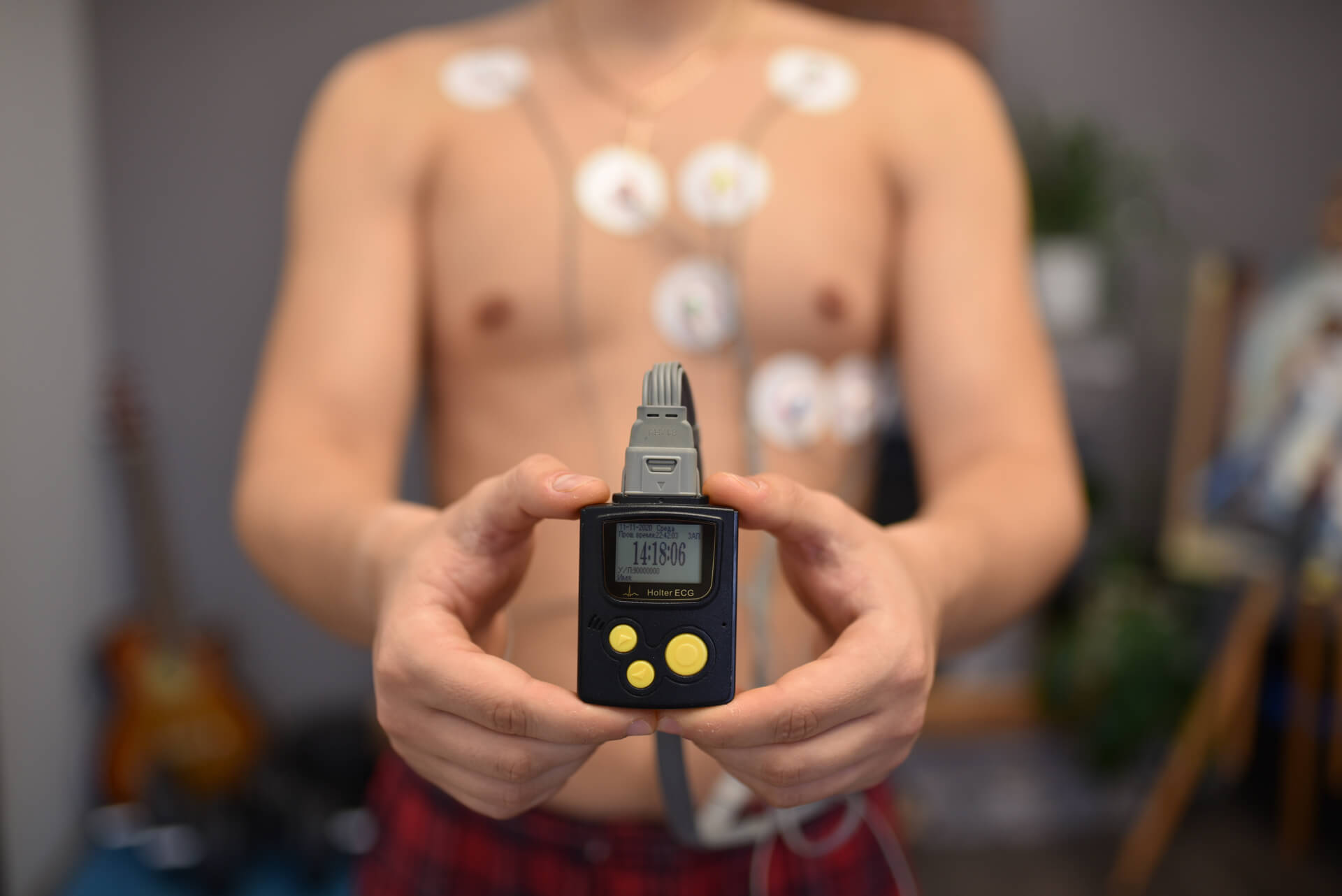
Heart Rhythm Monitoring
Prolonged heart rhythm monitoring is often required to investigate palpitations, dizziness or blackouts. This is usually done as a 24-hour or 48-hour Holter monitor or as a 7-day event recorder. Again, this test is non-invasive and painless and we expect you to perform normal activities during the test. It involves sticking 3 ECG stickers to the chest which are connected to a portable monitor that can be clipped to your belt. Once the monitoring period is complete the device is returned for detailed heart rhythm analysis. If an arrhythmia is identified the test can be repeated to assess for improvement with treatment.
Prolonged heart rhythm monitoring is often required to investigate palpitations, dizziness or blackouts. This is usually done as a 24-hour or 48-hour Holter monitor or as a 7-day event recorder. Again, this test is non-invasive and painless and we expect you to perform normal activities during the test. It involves sticking 3 ECG stickers to the chest which are connected to a portable monitor that can be clipped to your belt. Once the monitoring period is complete the device is returned for detailed heart rhythm analysis. If an arrhythmia is identified the test can be repeated to assess for improvement with treatment.

24hr Blood Pressure Monitoring
Blood pressure monitoring over a 24-hour period allows for more accurate measurement of your blood pressure. This has important implications when considering initiation of blood pressure medication and/or monitoring progress. This test is also non-invasive and painless. It involves placing a blood pressure cuff around the arm which automatically measures blood pressure several times per hour. We expect to perform normal activities during the test period. Once completed the device is returned for detailed blood pressure analysis.
Blood pressure monitoring over a 24-hour period allows for more accurate measurement of your blood pressure. This has important implications when considering initiation of blood pressure medication and/or monitoring progress. This test is also non-invasive and painless. It involves placing a blood pressure cuff around the arm which automatically measures blood pressure several times per hour. We expect to perform normal activities during the test period. Once completed the device is returned for detailed blood pressure analysis.
Exercise Stress Testing
It Is performed to evaluate your hearts behaviour under the physical stress of exercise. Typically this is performed to assess for coronary artery disease, however, it can also be performed to evaluate for heart rhythm disturbances, heart muscle disease or to assess the severity of heart valve disease. The test is non-invasive and painless. During the test you are asked to remove the top half of your clothing and are attached to an ECG machine and blood pressure recorder. You will be supervised by a trained cardiac physiologist throughout the test and recovery phase. Once you start on the exercise machine (treadmill or bicycle) the speed and intensity increase every three minutes. The test is completed when you achieve a target heart rate or if you develop symptoms that prevent you from carrying on. If you are unable to exercise the test can be done by artificially ‘stressing’ the heart with medication. You can go home after a brief recovery period (15-30 mins). After the test the heart rate, rhythm, blood pressure and ECG tracings are analysed for signs of heart disease and results forwarded to your Cardiologist.
It Is performed to evaluate your hearts behaviour under the physical stress of exercise. Typically this is performed to assess for coronary artery disease, however, it can also be performed to evaluate for heart rhythm disturbances, heart muscle disease or to assess the severity of heart valve disease. The test is non-invasive and painless. During the test you are asked to remove the top half of your clothing and are attached to an ECG machine and blood pressure recorder. You will be supervised by a trained cardiac physiologist throughout the test and recovery phase. Once you start on the exercise machine (treadmill or bicycle) the speed and intensity increase every three minutes. The test is completed when you achieve a target heart rate or if you develop symptoms that prevent you from carrying on. If you are unable to exercise the test can be done by artificially ‘stressing’ the heart with medication. You can go home after a brief recovery period (15-30 mins). After the test the heart rate, rhythm, blood pressure and ECG tracings are analysed for signs of heart disease and results forwarded to your Cardiologist.


Echocardiology
Uses ultrasound waves to evaluate the heart similar to scans performed for pregnant women. The scan is non-invasive and painless. You will be required to remove the top half of your clothing and will be hooked up to an ECG machine at the same time. Special ultrasound gel is used on the chest to improve the quality of the scan and this is usually performed in a darkened room by a trained cardiac physiologist. The echo will take 15-45 minutes and allows detailed evaluation of the heart valves and chambers and provides important information on structure and function. The echo also provides information regarding coronary artery disease, heart failure, heart muscle disease and heart valve disease if present. Specialist types of echo include contrast echo, stress echo and transesophageal echo that may be required in specific circumstances.
Uses ultrasound waves to evaluate the heart similar to scans performed for pregnant women. The scan is non-invasive and painless. You will be required to remove the top half of your clothing and will be hooked up to an ECG machine at the same time. Special ultrasound gel is used on the chest to improve the quality of the scan and this is usually performed in a darkened room by a trained cardiac physiologist. The echo will take 15-45 minutes and allows detailed evaluation of the heart valves and chambers and provides important information on structure and function. The echo also provides information regarding coronary artery disease, heart failure, heart muscle disease and heart valve disease if present. Specialist types of echo include contrast echo, stress echo and transesophageal echo that may be required in specific circumstances.
CT Coronary Angiography
Is used to evaluate the coronary arteries supplying the heart muscle with blood. Specifically, this is used to assess for narrowing or blockages of these arteries that may cause angina and lead to a heart attack. To perform the test a ‘cannula’ is inserted to a vein in your arm. This allows injection of special contrast ‘dye’ whilst you lie in a CT scanner. The test is performed by a trained radiographer under the supervision of a radiologist who will analyse the results and usually takes 15-20 minutes. A calcium score can also be taken of the coronary arteries (or the aortic valve) which can indicate disease in these areas. After the test you will be able to go home the same day after a short period of monitoring.
Is used to evaluate the coronary arteries supplying the heart muscle with blood. Specifically, this is used to assess for narrowing or blockages of these arteries that may cause angina and lead to a heart attack. To perform the test a ‘cannula’ is inserted to a vein in your arm. This allows injection of special contrast ‘dye’ whilst you lie in a CT scanner. The test is performed by a trained radiographer under the supervision of a radiologist who will analyse the results and usually takes 15-20 minutes. A calcium score can also be taken of the coronary arteries (or the aortic valve) which can indicate disease in these areas. After the test you will be able to go home the same day after a short period of monitoring.


Cardiac MRI Imaging
Allows detailed evaluation of the heart using an MRI scanner and can be used to diagnose and investigate heart muscle disease, heart failure, heart valve disease and damage to the heart after a heart attack. It can also be used in cases of congenital heart defects or where inflammation of the heart muscle or heart lining is suspected. The cardiac MRI requires you to lie still in a scanner for up to an hour and the machine is relatively noisy. You will be able to communicate with a trained radiographer during the scan at all times. Some people complain of claustrophobia during the scan and we will be able to support you through this. After the scan you will be able to go home the same day after a short period of monitoring.
Allows detailed evaluation of the heart using an MRI scanner and can be used to diagnose and investigate heart muscle disease, heart failure, heart valve disease and damage to the heart after a heart attack. It can also be used in cases of congenital heart defects or where inflammation of the heart muscle or heart lining is suspected. The cardiac MRI requires you to lie still in a scanner for up to an hour and the machine is relatively noisy. You will be able to communicate with a trained radiographer during the scan at all times. Some people complain of claustrophobia during the scan and we will be able to support you through this. After the scan you will be able to go home the same day after a short period of monitoring.
Implantable Loop Recorder
May be considered in cases where prolonged heart rhythm monitoring is required or if symptoms of palpitations, dizziness or blackouts are intermittent and/or infrequent. This is an invasive, day-case procedure performed in the Cardiac Catheter Lab under sterile conditions. A small local anaesthetic injection is administered on the left side of the chest and small incision made (5-8mm in length). A monitoring device is implanted under the skin and the wound closed. The device can stay in place for a few years if needed and data stored on the device can be downloaded and analysed for any evidence of arrhythmia.
May be considered in cases where prolonged heart rhythm monitoring is required or if symptoms of palpitations, dizziness or blackouts are intermittent and/or infrequent. This is an invasive, day-case procedure performed in the Cardiac Catheter Lab under sterile conditions. A small local anaesthetic injection is administered on the left side of the chest and small incision made (5-8mm in length). A monitoring device is implanted under the skin and the wound closed. The device can stay in place for a few years if needed and data stored on the device can be downloaded and analysed for any evidence of arrhythmia.


Invasive Coronary Angiography
Is the gold standard test to evaluate the coronary arteries in individuals with angina and suspected coronary artery disease. If you need this procedure you will undergo a pre-assessment by our specialist nursing team. This is an invasive, day-case procedure performed in the Cardiac Catheter Lab under sterile conditions. The procedure is done at the wrist with a local anaesthetic injection. A plastic ‘sheath’ is inserted into the right radial artery and plastic ‘catheters’ are passed to the heart under x-ray visualisation. Once at the heart, special ‘dye’ is injected into the coronary arteries and a series of images taken to evaluate the coronary arteries for narrowing or blockages. At this time, physiological evaluation of blood flow in the coronary arteries and/or direct imaging of the inside of the coronary vessels can be performed if clinically indicated. If coronary angioplasty is required, this can be performed at the same time. Afterwards you will be monitored for a few hours and discharged the same day.
Is the gold standard test to evaluate the coronary arteries in individuals with angina and suspected coronary artery disease. If you need this procedure you will undergo a pre-assessment by our specialist nursing team. This is an invasive, day-case procedure performed in the Cardiac Catheter Lab under sterile conditions. The procedure is done at the wrist with a local anaesthetic injection. A plastic ‘sheath’ is inserted into the right radial artery and plastic ‘catheters’ are passed to the heart under x-ray visualisation. Once at the heart, special ‘dye’ is injected into the coronary arteries and a series of images taken to evaluate the coronary arteries for narrowing or blockages. At this time, physiological evaluation of blood flow in the coronary arteries and/or direct imaging of the inside of the coronary vessels can be performed if clinically indicated. If coronary angioplasty is required, this can be performed at the same time. Afterwards you will be monitored for a few hours and discharged the same day.
